Panoramic X-rays and CT scans
Understanding Panoramic X-rays and CT Scans: Essential Tools for Dental Health
In the realm of dental health, precision and clarity are paramount, and advancements in imaging technology have revolutionized the way oral care professionals diagnose and treat patients. Among these innovations, panoramic X-rays and CT scans stand out as essential tools that provide comprehensive views of the mouth, teeth, and jaws. These imaging techniques offer invaluable insights, allowing dentists to detect underlying issues that may not be visible through traditional methods. Whether you’re preparing for a routine check-up or facing a more complex dental procedure, understanding the differences and applications of these imaging modalities can empower you to make informed decisions about your oral health. In this blog post, we will delve into the intricacies of panoramic X-rays and CT scans, exploring how they work, what they reveal, and why they are crucial in maintaining optimal dental health for patients of all ages. Join us as we demystify these essential tools and highlight their role in ensuring a healthier smile for you and your loved ones.
1. Introduction to Panoramic X-rays and CT Scans
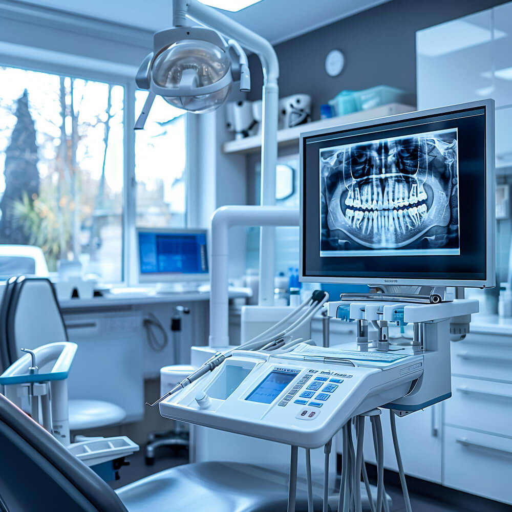
In the realm of dental health, the ability to visualize the complex structures of the mouth and jaw is paramount for accurate diagnosis and effective treatment planning. Among the advanced imaging techniques available to dentists, panoramic X-rays and CT scans stand out as essential tools that provide a comprehensive view of oral health.
Panoramic X-rays, often referred to as orthopantomograms (OPGs), capture a single, two-dimensional image that encompasses the entire mouth, including the teeth, jaw, and surrounding structures. This unique perspective allows dental professionals to assess the alignment of teeth, diagnose cavities, evaluate bone density, and identify potential issues such as impacted teeth or jaw disorders. The convenience of panoramic X-rays lies in their ability to deliver a broad overview in a single exposure, making them an invaluable resource for routine check-ups and initial evaluations.
On the other hand, CT scans—specifically cone beam computed tomography (CBCT)—offer a more detailed, three-dimensional view of the dental and skeletal structures. This advanced imaging technique is particularly beneficial for complex cases, such as dental implants, orthodontic assessments, and the evaluation of cysts or tumors. CBCT scans provide high-resolution images that reveal intricate anatomical details, enabling dentists to make more informed decisions and tailor treatment plans to meet the specific needs of each patient.
As we delve deeper into the world of panoramic X-rays and CT scans, it’s essential to understand their unique benefits, applications, and the role they play in enhancing diagnostic accuracy and improving overall dental care. By leveraging these innovative imaging technologies, dental professionals can ensure that their patients receive the highest standard of care in maintaining and restoring their oral health.
2. What Are Panoramic X-rays?
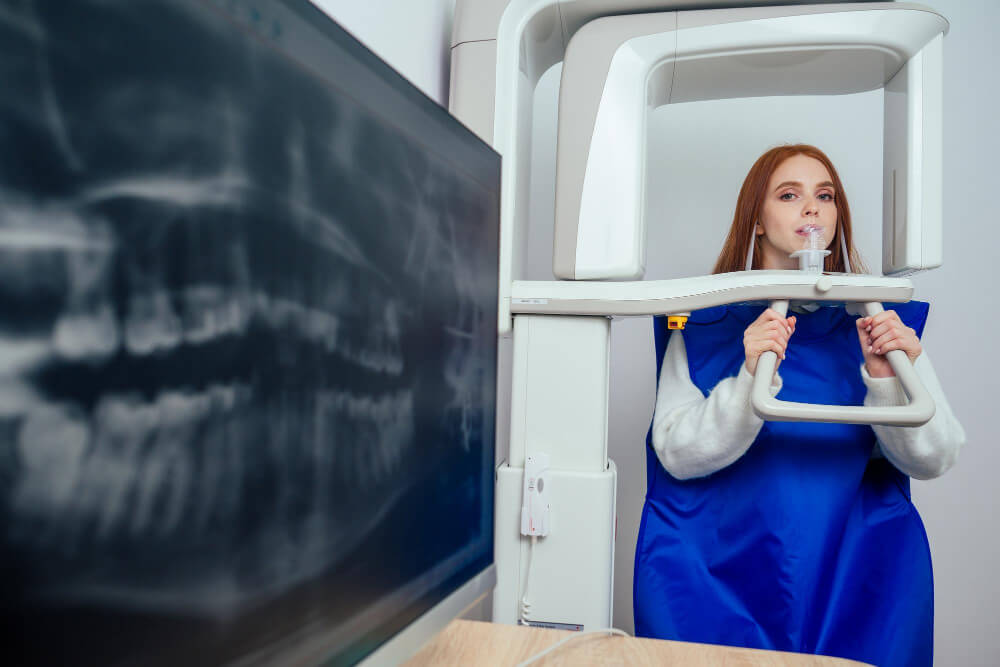
Panoramic X-rays, also known as panoramic radiographs, are a specialized type of dental imaging that captures a comprehensive view of the entire mouth in a single, sweeping shot. Unlike traditional X-rays, which focus on individual teeth or specific sections of the mouth, panoramic X-rays provide a broad overview, encompassing the upper and lower jaws, teeth, sinuses, and even the temporomandibular joints (TMJ). This wide-angle perspective is invaluable for dentists as it allows them to assess overall oral health, evaluate tooth alignment, and identify potential issues that may not be visible through standard X-rays.
The process of obtaining a panoramic X-ray is quick and straightforward. Patients stand comfortably in front of the machine as it rotates around their head, capturing images from different angles. This method not only minimizes exposure to radiation—making it a safer option—but also vastly reduces the time and effort required for multiple individual X-rays.
One of the key benefits of panoramic X-rays is their ability to reveal important information about dental structures that may be hidden beneath the surface. For example, they can help detect impacted teeth, such as wisdom teeth, identify jaw abnormalities, and assess the condition of bony structures. This comprehensive view aids dentists in developing effective treatment plans, whether it’s for routine care, orthodontics, or surgical interventions.
Overall, panoramic X-rays are an essential tool in modern dentistry, offering a strategic advantage in diagnosing and monitoring dental health. By providing a holistic view of the mouth, they empower dental professionals to make informed decisions and ultimately enhance patient care.
3. How Panoramic X-rays Work
Panoramic X-rays, often referred to as panorex or panoramic radiographs, provide a broad view of the entire mouth in a single image. Unlike traditional X-rays that focus on specific areas, panoramic X-rays capture the full landscape of the dental and facial structures, offering invaluable insights into oral health.
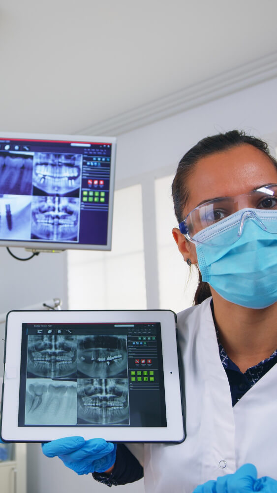
The process begins with the patient standing or sitting in front of the X-ray machine, which resembles a large, circular apparatus. As the machine rotates around the patient’s head, it emits a low dose of radiation to capture images from multiple angles. This rotation is what allows for a comprehensive view, effectively creating a two-dimensional image that displays the teeth, gums, jawbone, and even the sinuses and temporomandibular joints (TMJ) in one frame.
The technology behind panoramic X-rays utilizes a film or digital sensor that records the images as the machine moves. The resulting image is a continuous view of the dental arches, showing not only the positioning of the teeth but also the alignment of the jaw and any potential issues lurking below the surface. This capability is particularly beneficial for orthodontic assessments, wisdom tooth evaluations, and detecting jaw abnormalities or cysts.
One of the key advantages of panoramic X-rays is their ability to reveal problems that may not be visible in standard X-rays. For instance, they can highlight impacted teeth, bone loss, or tumors, providing a more complete picture of a patient’s oral health. Moreover, the process is quick, typically taking less than a minute, with minimal discomfort for the patient.
By understanding how panoramic X-rays work, patients can appreciate their significance in preventive care and early diagnosis, setting the stage for more effective treatment plans and better overall dental health.
4. Benefits of Panoramic X-rays in Dentistry
Panoramic X-rays, also known as orthopantomograms, play a pivotal role in modern dentistry, offering a comprehensive view of a patient’s oral health with a single image. The primary advantage of panoramic X-rays lies in their ability to capture a broad view of the entire mouth, including the upper and lower jaws, teeth, and surrounding structures, in one seamless shot. This panoramic approach allows dentists to assess various aspects of dental health that might be missed with traditional X-rays.
One significant benefit is the ability to detect issues early. Panoramic X-rays can reveal impacted teeth, such as wisdom teeth that may not have erupted properly, as well as cysts and tumors that could indicate more serious conditions. By identifying these problems early, dentists can formulate timely and effective treatment plans, potentially preventing more invasive procedures down the line.
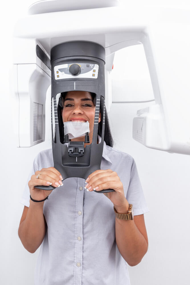
Additionally, the panoramic X-ray process is quick and painless. Patients stand in one spot and the machine rotates around their head, capturing the full view in about 10-20 seconds. This efficiency not only enhances patient comfort but also streamlines the workflow in dental practices, ensuring that patients spend less time waiting and more time receiving care.
Another important aspect of panoramic X-rays is their ability to assist in treatment planning for orthodontics and implantology. Orthodontists can use these images to evaluate jaw relationships and tooth alignment, while oral surgeons can assess bone structure and density before placing implants. This comprehensive view is instrumental in achieving successful outcomes and minimizing complications.
Lastly, panoramic X-rays are invaluable for fostering communication between dentists and patients. The clear visual representation of dental structures allows dentists to explain diagnoses and treatment plans more effectively, helping patients to understand their oral health better and encouraging them to take an active role in their care.
In summary, the benefits of panoramic X-rays in dentistry are multi-faceted, providing early detection of dental issues, enhancing treatment planning, streamlining procedures, and improving patient communication—all essential components in maintaining optimal dental health.
5. What Are CT Scans?
CT scans, or computed tomography scans, are advanced imaging techniques that provide detailed cross-sectional images of the teeth, jaw, and surrounding structures. Unlike traditional X-rays, which capture a two-dimensional view, CT scans create a three-dimensional representation of the oral cavity, allowing dental professionals to observe complex anatomy with remarkable clarity. This technology utilizes a series of X-ray images taken from various angles, which are then processed by a computer to produce comprehensive images that reveal even the smallest details.
One of the primary advantages of CT scans in dentistry is their ability to aid in diagnosing conditions that might not be visible through standard X-rays. For instance, they are invaluable in the assessment of dental implants, helping practitioners evaluate bone density and structure before surgery. Additionally, CT scans can uncover issues such as tumors, cysts, and impacted teeth, providing a clear visual roadmap for effective treatment planning.
The precision of CT imaging also extends to evaluating the surrounding anatomical features, such as the sinuses and nerves, which is crucial for procedures like wisdom tooth extractions or complex orthodontic treatments. This comprehensive overview helps dentists devise tailored treatment strategies, ensuring optimal outcomes for patients.
While the benefits of CT scans are significant, it’s essential to note that they do involve a higher level of radiation exposure compared to traditional X-rays. Therefore, dental professionals typically reserve CT scans for specific cases where detailed imaging is necessary to enhance patient care. With their ability to reveal intricate details of dental anatomy, CT scans stand as an essential tool in modern dentistry, empowering practitioners to make informed decisions and deliver superior care to their patients.

6. How CT Scans Differ from Traditional X-rays
When it comes to dental imaging, understanding the differences between CT scans and traditional X-rays is crucial for both patients and practitioners. While both technologies serve the purpose of visualizing the internal structures of the mouth and jaw, their methodologies and the depth of information they provide vary significantly.
Traditional X-rays, or two-dimensional images, capture a flat picture of the dental anatomy. They are typically used for diagnosing common issues, such as cavities, impacted teeth, and bone loss. However, the limitations of traditional X-rays become apparent when it comes to complex cases. For instance, overlapping structures can obscure important details, making it difficult for dentists to assess the precise condition of teeth and surrounding tissues.
On the other hand, Computed Tomography (CT) scans offer a revolutionary leap in imaging technology. CT scans utilize a series of X-ray images taken from different angles and then compile them into a comprehensive three-dimensional view of the dental structures. This allows for a far more detailed examination of the bones, soft tissues, and even the nerves within the jaw. With this 3D perspective, dental professionals can identify issues that might go unnoticed in traditional X-rays, such as subtle fractures, bone abnormalities, and complex root canal formations.
Moreover, the increased accuracy of CT scans can lead to more effective treatment planning, particularly for complex procedures like dental implants or surgeries. They enable dentists to visualize the exact anatomy of the area being treated, which can significantly improve outcomes and patient safety.
In summary, while traditional X-rays provide a quick and accessible snapshot of dental health, CT scans open up a new realm of diagnostic possibilities. By offering detailed, three-dimensional images, CT scans equip dentists with the essential tools they need to provide comprehensive care and ensure optimal outcomes for their patients.
7. Advantages of CT Scans in Dental Diagnosis
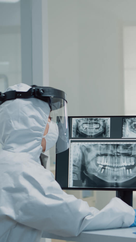
CT scans, or computed tomography scans, have revolutionized the landscape of dental diagnosis, offering a wealth of advantages that enhance both the precision of assessments and the quality of patient care. Unlike traditional X-rays, which provide flat, two-dimensional images, CT scans create detailed, three-dimensional representations of the oral and maxillofacial regions. This capability allows dental professionals to visualize complex anatomical structures with unprecedented clarity.
One of the standout advantages of CT scans is their ability to detect issues that may be overlooked by conventional methods. For instance, they excel in identifying hidden dental pathologies, such as bone loss, cysts, or tumors, allowing for early intervention and more effective treatment planning. This is particularly beneficial in cases of dental implants, where precise measurements of bone density and structure are crucial for success.
Moreover, the comprehensive nature of CT imaging aids in diagnosing temporomandibular joint (TMJ) disorders and sinus issues related to dental health. By providing a holistic view of the jaw, teeth, and surrounding tissues, CT scans empower dentists to devise more tailored treatment strategies, ultimately leading to better patient outcomes.
Additionally, the speed and efficiency of CT scans make them an invaluable tool in emergency dental situations. When time is of the essence, such as in trauma cases, the quick acquisition of detailed images can be lifesaving. This rapid response capability allows dental professionals to make informed decisions swiftly, ensuring that patients receive the appropriate care without unnecessary delays.
Overall, the advantages of CT scans in dental diagnosis are manifold. They not only enhance the accuracy of assessments but also facilitate more effective treatment planning, improve patient management, and ultimately elevate the standard of dental care. As technology continues to advance, incorporating CT scans into routine dental practice will undoubtedly play a pivotal role in safeguarding and improving dental health.
8. When Are Panoramic X-rays Used?
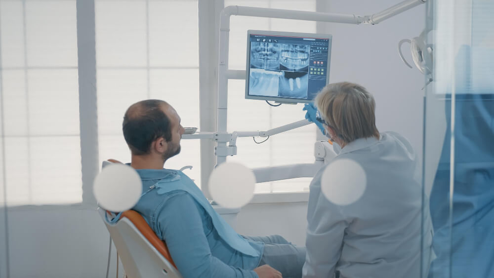
Panoramic X-rays are a vital tool in modern dentistry, offering a comprehensive view of a patient’s oral health in a single image. Unlike traditional X-rays, which focus on specific teeth or areas, panoramic X-rays capture the entire dental arch, including the upper and lower jaws, teeth, and surrounding structures. This broad perspective allows dentists to assess a range of conditions and plan effective treatments.
So, when exactly are panoramic X-rays used? They are particularly beneficial in several scenarios:
1. **Initial Assessments**: During a patient’s first visit, panoramic X-rays can provide a baseline overview of their oral health. Dentists can identify potential issues, such as impacted teeth, jaw abnormalities, or signs of periodontal disease that may not be visible during a standard examination.
2. **Orthodontic Treatment Planning**: For patients considering braces or other orthodontic interventions, panoramic X-rays are indispensable. They reveal the position of teeth, including those that have yet to erupt, and help orthodontists devise a tailored treatment plan.
3. **Wisdom Tooth Evaluation**: When assessing the need for wisdom tooth extraction, panoramic X-rays are the go-to choice. They can show the position of the wisdom teeth relative to the surrounding teeth and nerves, informing the surgeon about the best approach to extraction.
4. **Dental Implants**: For patients seeking dental implants, panoramic X-rays provide critical information about bone density and structure. This data is essential for determining the feasibility of implant placement and ensuring the long-term success of the procedure.
5. **Trauma Assessment**: In cases of dental trauma, such as fractures or dislocations, panoramic X-rays allow dentists to visualize the extent of the injury across the entire jaw, aiding in accurate diagnosis and treatment planning.
By utilizing panoramic X-rays in these various scenarios, dental professionals can enhance their diagnostic capabilities and ensure that patients receive the most effective care tailored to their unique needs. This holistic approach not only improves treatment outcomes but also fosters a deeper understanding of each patient’s dental health journey.

9. When Are CT Scans Recommended?
CT scans, or computed tomography scans, are advanced imaging techniques that provide detailed cross-sectional images of the teeth, jaw, and surrounding structures. While traditional X-rays are often sufficient for routine examinations, there are specific scenarios where a CT scan is recommended for a more comprehensive view of dental health.
1. **Complex Dental Issues**: If a patient presents with complicated dental problems, such as impacted teeth or extensive decay that may require surgical intervention, a CT scan can offer invaluable insights. It allows dentists to visualize the precise location and orientation of the teeth and roots, aiding in accurate diagnosis and treatment planning.
2. **Implant Planning**: For patients considering dental implants, CT scans are essential. They help in assessing the bone density and structure where the implant will be placed, ensuring that there is adequate bone and identifying any anatomical variations that could affect the procedure. This information is crucial for achieving a successful implant placement.
3. **Jaw Disorders**: Conditions like temporomandibular joint (TMJ) disorders can be challenging to diagnose with standard X-rays. A CT scan provides a clear view of the jaw joints, enabling dentists to assess the bone structure and any abnormalities that could be contributing to pain or dysfunction.
4. **Oral Pathology**: If there are signs of abnormal growths or lesions in the oral cavity, a CT scan can help determine the extent of the issue. This imaging modality allows for a more thorough examination of the surrounding tissues, assisting in the diagnosis of conditions such as cysts, tumors, or infections.
5. **Trauma Assessment**: In cases of dental trauma, where fractures or dislocations may occur, a CT scan is often recommended. It can provide a detailed look at the extent of the injury, allowing for more effective treatment planning.
6. **Diagnosis of Sinus Issues**: Dental health is closely linked to sinus health, particularly in the upper jaw area. A CT scan can reveal sinus infections or other issues that may be contributing to dental pain or discomfort, helping dentists to treat the root cause effectively.
In summary, while CT scans are not typically the first line of imaging for every dental issue, they are invaluable in complex cases that require a deeper understanding of a patient’s dental and oral health. By providing detailed images that reveal the intricacies of the jaw and teeth, CT scans empower dental professionals to make informed decisions and deliver tailored treatments, ultimately enhancing patient outcomes.
10. Understanding the Safety of X-rays and CT Scans
When it comes to dental health, the safety of X-rays and CT scans is a common concern for patients. Understanding the risks and benefits associated with these diagnostic tools is essential for making informed decisions about your oral care.
Dental X-rays, which have been in use for over a century, emit very low doses of radiation—approximately 0.5 to 5 microsieverts per image, depending on the type of X-ray taken. To put this in perspective, this amount is comparable to the natural background radiation a person is exposed to over just a few days. Furthermore, advancements in dental imaging technology have continuously reduced radiation exposure, meaning that modern X-ray machines are safer than ever before.
CT scans, or computed tomography scans, are more advanced imaging techniques that provide detailed three-dimensional images of the dental structures and surrounding tissues. While they do expose patients to higher levels of radiation than traditional X-rays, the information gained can be crucial for diagnosing complex dental issues, such as impacted teeth or jawbone abnormalities. It’s important to note that the benefits of obtaining a clear and comprehensive view of dental health often outweigh the risks posed by the radiation exposure.
Dental professionals prioritize patient safety and are guided by the “As Low As Reasonably Achievable” (ALARA) principle, which emphasizes minimizing radiation exposure while still obtaining the necessary diagnostic information. They carefully evaluate the need for imaging based on individual patient circumstances, ensuring that X-rays and CT scans are only performed when essential for effective treatment planning.
Before undergoing any imaging procedure, patients should not hesitate to ask their dentist about the safety measures in place, the necessity of the scans, and the potential risks involved. Open communication can help alleviate concerns and foster a better understanding of the role these diagnostic tools play in maintaining optimal dental health. Ultimately, when performed judiciously and with the right precautions, X-rays and CT scans are invaluable tools that contribute significantly to effective dental care.
11. Preparing for Your Panoramic X-ray or CT Scan
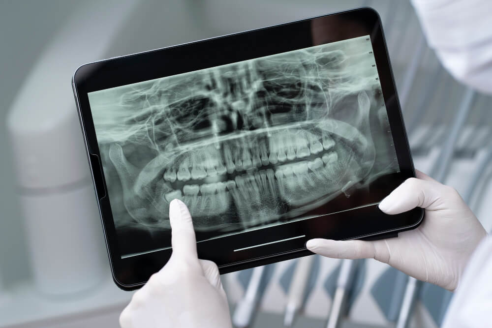
Preparing for your panoramic X-ray or CT scan is a crucial step that ensures the procedure goes smoothly and yields the most accurate results. Before your appointment, it’s essential to communicate openly with your dental professional about any medical conditions or allergies you may have. This information can help them tailor the procedure to your specific needs and ensure your safety.
On the day of your appointment, you’ll want to wear comfortable clothing without any metal fasteners, such as zippers or buttons, which can interfere with the imaging process. It’s advisable to leave jewelry, eyeglasses, and any other accessories at home, as these items can create artifacts on the X-ray images.
As you arrive at the dental office, you may be asked to sign consent forms and review your medical history once more. This is also a great time to ask any questions you have about the procedure itself—understanding the steps involved can help ease any anxiety you may feel.
Once you’re in the imaging room, the technician will position you appropriately, ensuring that your head is stable and aligned. For a panoramic X-ray, you’ll typically bite down on a small, comfortable device to keep your mouth in the correct position. If you’re undergoing a CT scan, you may need to lie down on a table that moves through the CT machine. During the scan, it’s important to remain as still as possible for the best image quality.
Finally, if your dentist has recommended a contrast dye for your CT scan, you might receive instructions on how to prepare for it, such as fasting for a few hours beforehand. Following these guidelines will help ensure that your panoramic X-ray or CT scan provides the detailed images necessary for accurate diagnosis and treatment planning, paving the way for optimal dental health.

12. What to Expect During the Procedure
When it comes to dental imaging, both panoramic X-rays and CT scans play crucial roles in diagnosing and planning treatments. Understanding what to expect during these procedures can help ease any anxiety and ensure a smooth experience.
**Panoramic X-rays:**
Before your panoramic X-ray, your dentist will explain the process and what the images will reveal about your dental health. You’ll be asked to remove any jewelry or accessories that might interfere with the imaging.
Once you’re ready, you’ll stand in front of the X-ray machine, which resembles a large, circular device. A bite block will be placed in your mouth to help you maintain the correct position, while the machine rotates around your head, capturing a comprehensive view of your teeth, jaw, and surrounding structures. The entire process typically takes less than a minute, and you’ll be required to stay still while the images are being taken. Afterward,
the X-ray images will be reviewed by your dentist, who will discuss their findings and recommend any necessary treatments.
**CT Scans:**
A CT scan, or computed tomography scan, is slightly more complex but still relatively straightforward. Before the procedure, your dentist will again explain the process. You’ll be asked to remove any metal objects, such as hairpins or glasses, and you may be positioned in a reclining chair or on a table that moves through the scanning machine. Unlike a panoramic X-ray, a CT scan takes multiple images from different angles, which are then compiled by a computer to create detailed cross-sectional views of your teeth, gums, and bone structure.
The scan itself usually takes about 30 seconds to a few minutes, but you may be in the room for a bit longer due to preparation and positioning. The procedure is painless, and you’ll be able to resume your normal activities immediately afterward.
Both procedures are non-invasive and designed to provide your dentist with crucial information to help maintain your dental health. By knowing what to expect, you can approach your appointment with confidence, ensuring a positive experience that contributes to your overall well-being.
13. Interpreting the Results: What Do They Mean?
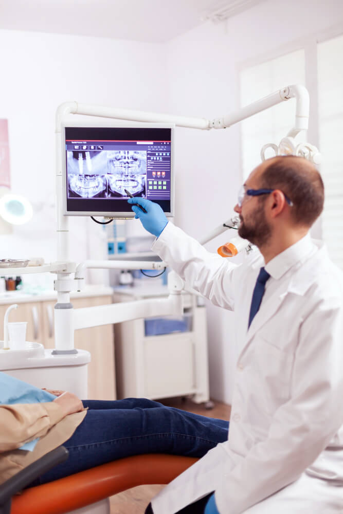
Interpreting the results of panoramic X-rays and CT scans is a crucial step in understanding your dental health. These imaging techniques provide a comprehensive view of your mouth, teeth, and surrounding structures, revealing details that are not visible through standard dental exams. However, translating these images into actionable insights requires a keen eye and an understanding of what the various findings indicate.
**Panoramic X-rays**, for instance, deliver a two-dimensional view of the entire mouth in a single image. This broad perspective allows dentists to assess the overall alignment of teeth, the presence of impacted wisdom teeth, or any anomalies in jaw structure. When reviewing these X-rays, your dentist will look for signs of decay, periodontal disease, and other dental issues that may require attention. Each shadow and contour tells a story, and your dentist will explain what each of these elements means in the context of your oral health.
On the other hand, **CT scans** offer a more detailed, three-dimensional view, making it easier to identify complex conditions such as jaw fractures, tumors, or cysts. The high-resolution images produced by CT scans help in diagnosing issues with unprecedented accuracy. Your dentist will examine these scans for specific indicators, such as the bone density around your teeth or the proximity of dental roots to critical structures like nerves and sinuses. Understanding these results is essential, as they can directly influence treatment planning, whether it’s a simple filling, orthodontic work, or more complex surgical procedures.
When discussing the results with your dental professional, don’t hesitate to ask questions. Understanding the implications of what you see on the screen can empower you to make informed decisions about your treatment options. This collaborative approach not only enhances your awareness of your dental health but also fosters a stronger relationship between you and your dental care provider. Ultimately, making sense of these imaging results is about more than just numbers and images; it’s about ensuring you receive the best possible care for your unique dental needs.

14. The Role of Panoramic X-rays and CT Scans in Treatment Planning
When it comes to effective treatment planning in dentistry, panoramic X-rays and CT scans play a pivotal role in providing comprehensive insights into a patient’s oral health. These imaging techniques are not just tools for diagnosis; they are integral to formulating an individualized treatment strategy that addresses each patient’s unique needs.
Panoramic X-rays offer a broad view of the entire mouth in a single image, capturing essential details about the teeth, jaws, and surrounding structures. This wide-angle perspective allows dentists to identify potential issues such as impacted teeth, jaw alignment problems, and the overall condition of bone structures. With this valuable information at hand, dental professionals can pinpoint areas that may require intervention, whether it be routine procedures like fillings or more complex treatments such as extractions or orthodontics.
On the other hand, CT scans provide a three-dimensional view that takes diagnostic precision to the next level. This advanced imaging technique allows for a detailed visualization of the bone density and structure, which is particularly beneficial in planning treatments for dental implants and complex surgical procedures. By evaluating the intricate anatomy of the jaw and surrounding tissues, dentists can make informed decisions about implant placement, anticipate potential complications, and customize surgical approaches to enhance patient outcomes.
Moreover, the integration of panoramic X-rays and CT scans in treatment planning fosters collaborative discussions among dental specialists. For instance, oral surgeons, orthodontists, and periodontists can share insights and devise a cohesive treatment plan that addresses all aspects of a patient’s dental health. This multidisciplinary approach not only optimizes the effectiveness of treatments but also ensures that the patient receives comprehensive care tailored specifically to their needs.
In conclusion, the role of panoramic X-rays and CT scans in treatment planning cannot be overstated. These imaging technologies serve as essential tools that empower dental professionals to diagnose accurately, plan effectively, and ultimately provide patients with the highest standard of care. By embracing these advanced techniques, dentists can enhance their ability to deliver successful outcomes, ensuring that patients leave with healthy smiles and increased confidence in their oral health journey.
15. Conclusion: Choosing the Right Imaging Tool for Dental Health
In conclusion, selecting the right imaging tool for dental health is a crucial decision that can significantly impact both diagnosis and treatment outcomes. Panoramic X-rays and CT scans each offer unique advantages tailored to specific dental needs. Panoramic X-rays provide a comprehensive view of the entire mouth, capturing essential information about the teeth, jaw, and surrounding structures in a single, quick image. This makes them an excellent choice for routine examinations, orthodontic assessments, and pre-surgical planning. Their lower radiation exposure compared to other imaging methods is an added bonus, making them suitable for all patients, including children.
On the other hand, CT scans offer unparalleled detail and precision, revealing intricate structures that may be missed by standard X-rays. This advanced imaging modality is particularly beneficial for complex cases, such as impacted teeth, jaw disorders, and the evaluation of tumors or cysts. However, the higher radiation dose and cost associated with CT scans necessitate careful consideration and should be reserved for situations where detailed cross-sectional images are essential for effective diagnosis and treatment planning.
Ultimately, the choice between these two imaging techniques should be guided by the specific dental scenario at hand, the level of detail required, and the patient’s health considerations. Consulting with a knowledgeable dental professional can help patients navigate their options, ensuring that they receive the most appropriate imaging that aligns with their unique dental health needs. By understanding the strengths and limitations of both panoramic X-rays and CT scans, patients can make informed choices that pave the way for optimal dental care and long-term oral health.
